Acute leukemia – is acute malignant disease of the blood system, with defeats at the level of deterministic genus-parent stem cells or early cellular predecessor, characterized by the availability of blast cells in the bone marrow puncture or in peripheral blood from 20 % and more.[4].
Today, at the diagnosis of acute myeloid leukemia by dint of flowing cytofluorimetry, it is necessary to appreciate the immunophenotypic features of tumor (blast) cells and define the directionality of myeloid cell linearity and important exclude T and B -linear or rare variants of unclear linearity [3].
Cells of the myeloid linearity develop in the bone marrow from a general hematopoietic predecessor. The early cells monocytic series – are monoblasts and promonocytes. In norm their content in the bone marrow is utterly low.
By description to the author [7], monoblasts express markers of the predecessors CD34, CD117 and HLA-DR, the appear in the sequence CD4, CD64, CD36, CD33, CD11c, CD11b, CD15, CD14, and in stage promonocyte the intensity expression of these given markers increases, and antigens of the early stage of differentiation (CD34, CD117) slowly disappear.
At the diagnosis of acute myeloid leukemia (AML), the most specific is the elicitation of myeloperoxidase (MPO) in the cytoplasm of tumor cells.
Myeloperoxidase-is a linear-specific marker of the myeloid line, a lysosomal enzyme granulocytes.
In immunophenotyping in the case of not detecting myeloperoxidase, that for establish the myeloid variant of acute leukemia, it is necessary to explore other myeloid antigens, including markers of rare forms of AML (erythroid, megakaryoblastic). With the diagnostic purposes the definition of the linear accessories of tumor cells uses sets of antibodies, recommended by an international group of experts [6], and for differential diagnosis, is using classifications the EGIL[5] and of the World Health Organization (WHO) [9].
For confirm the myeloid directionality of tumor (blast)cells, it is necessary to appreciate the expression of myeloid antigens.
By view to the authors [9], in acute myeloid leukemia (AML) blast cells most often express predecessor markers (CD34, CD117, HLA-DR) and in rare happening terminal deoxynucleotiddyl transferase (TdT), specialized DNK-polymerase, which in norm subjoin N-nucleotides at rearranging the T and B genes of cell receptors, increasing their variability.
At the acute myeloid leukemia marker СD7 detection in 30 % of cases, the expression of this antigen may correlate with not favorable prognosis.[8].
At making a diagnosis of myeloid option of linearity and correct selection of PChT, it is necessary to conduct allogeneic transplantation of hematopoietic stem cells. Timely conduct of a high-technology method of therapy, closely related, unrelated bone marrow transplantation, allows at the availability HLA – identical healthy donor [2], or placental blood.[1].
The aim of our study is to elicitation frequency the prevalence and timely diagnostic analysis of the immunophenotype of tumor (blast) cell linearity in acute myeloid leukemia in patients children in the Kyrgyz Republic(Kirgizia).
Materials and research methods
The group of research from November 2016 to December 2018 included 30 patients of children (female-16,male-14)with acute myeloid leukemia, of them patients children of kirgiz nationality-24 (female-14 and male-10) and residents of the Russian-speaking population of the Kyrgyz Republic (mixed nation and different nationalities) – 6 patients of children, of them ((female – 2, and male – 4), all citizens of the Kyrgyz Republic (Kirgizia), aged from 1.5 years to 16 years, who were examined at the Department of Pediatric Oncology of the National Center Oncology and Hematology Department of Health of the Kyrgyz Republic (Kirgizia) and in the Department of Pediatric Hematology Osh interregional clinical children’s hospital in Osh, in St. Petersburg consult doctor-hematologists Eurasian Center of Oncohematology, Immunology and Therapy.
Method immunophenotyping by dint of flowing cytofluorimetry conducted the first time and research immunophenotyping was conducted in Bishkek, Kyrgyz Republic(Kirgizia).[3].
Method by dint of flowing cytofluorimetry
The material for the study is the bone marrow. Patients made puncture of the sternum. For obtain a qualitative result, the obtained analysis should not be with impurity blood and not destroy the cells during the test-sample preparation to immunophenotyping. Immunophenotyping of leukemia (blast) cells performed on a flow cytofluometer Cytomics FC500 (Beckman Coulter, USA) using monoclonal antibodies Beckman Coulter.
Statistical processing of the results included the analysis of standard criteria X2-square, which was used to assess the significance of differences in the occurrence of certain characteristics between the control group and the test group. Determination of the “p”, the corresponding value found. X2-square performed considering one degree of freedom.
All mathematical calculations and statistical analysis of the overall study was performed using a personal computer using the package application programs for spreadsheets – Microsoft – Excel M version 7.0, for Windows 95, for Windows-based 2010, Statistica-5.
Research results and discussion
During our research, the diagnosis of acute myeloid leukemia was established on the basis of clinical data and a complex series of general and special laboratory-diagnostic indicators.

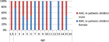
Fig. 1.Comparative age characteristics of the frequency of elicitation in the Kyrgyz Republic (Kirgizia) among patient’s children female and male with acute myeloid leukemia
By the results of our research as it is seen of the present in the Figure 1, in Kyrgyz Republic (Kirgizia) acute myeloid leukemia among patients children male at 1,5 years, 3 years, 8 years, 10 years and 14 years, and among patients female in 7 years, 9 years, 11 years and 13 years at these ages practically detection in 100 % of cases.
The detected among patients children females in the age of 2 years is detected among females in 77 % of cases, in 4 years in 50 % of cases, in 5 years in 32 % of cases, in 6 years in 50 % of cases and in 15 years in 50 % of cases and among patients children male at the age of 2 years in 23 % of cases, in 4 years in 50 % of cases, in 5 years in 68 % of cases, in 6 years in 50 % of cases, in 15 years in 50 % of cases.
At the our observation in the age of 12 years and 16 years, not elicitation among nor females, nor among males – 0 % of cases.
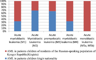
By the frequency prevalence among patients children kirgiz nationality is detected acute myeloblastic leukemia (variant M1) in 32 % of cases, acute myeloblastic leukemia (variant 2) in 69 % of cases, acute promyeloblastic leukemia(variant M3) in 67 % of cases, acute myeloblastic leukemia (variant M4) in 32 % of cases, acute monoblastic leukemia (variant M5a, M5b) in 0 % of cases, data are presented in Fig. 2.
In the compared among the patients children of residents of the Russian-speaking population of the Kyrgyz Republic (mixed nation and different nationalities)(Kirgizia), acute myeloblastic leukemia (variant M1) is detected in 68 % of cases, acute myeloblastic leukemia (variant 2) in 31 % of cases, acute promyeloblastic leukemia(variant M3) in 33 % of cases, acute myeloblastic leukemia (variant M4) in 68 % of cases, acute monoblastic leukemia (variant M5a, M5b) in 100 % of cases.
By of significance acute myeloblastic leukemia (variant M1), acute myeloblastic leukemia (variant M4), acute monoblastic leukemia (variant M5a, M5b), have statistically highly authentic differences, where, p < 0.0001.
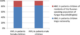
As can be seen from the presented Fig. 3, acute myeloid leukemia in female patients children of kirgiz nationality the elicitation in 78 % of cases, and among male patients in 83 % of cases.
And among patients children female of residents of the Russian-speaking population of the Kyrgyz Republic (Kirgizia) are detected in 22 % of cases, among males in 17 % of cases.
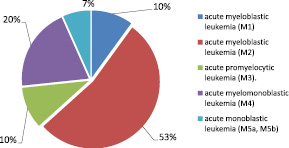
As can be seen from Fig. 4, is elicitation among the patients children of the Kyrgyz Republic, the most of the prevalence acute myeloblastic leukemia (M2 variant) in 53 % of cases, acute myelomonoblastic leukemia (M4 variant) in 20 % of cases and a little less spread acute promyelocytic leukemia (M3 variant) in 10 % of cases, acute myeloblastic leukemia (M1 variant) in 10 % of cases, acute monoblastic leukemia (5a,5b variant) in 7 % of cases.

Fig. 2. Variants of acute myeloid leukemia and the frequency of the prevalence among the patients children of kirgiz nationality and of the residents Russian-speaking population of the Kyrgyz Republic (Kirgizia)

Fig. 3. Prevalence among female and male patients children of acute myeloid leukemia in the Kyrgyz Republic (Kirgizia)

Fig. 4.The frequency of the prevalence in the Kyrgyz Republic among the patients children immunological option of acute myeloid leukemia
Thus, for determine the variant of linearity and elicitation frequency of prevalence, and the feature of the flow of acute myeloid leukemia in patient’s children in the Kyrgyz Republic, it is necessary conduct research by dint of flowing cytofluorimetry.
At the acute myeloid leukemia, despite in studying the detailed characteristics of tumor (blast) cells, which allows detection to the characteristic immunophenotype by dint of flowing cytofluorimetry, for reaffirm the diagnosis, it is necessary to conduct in parallel cytogenetic, molecular-genetic and morphological studies.
In the frequency of the prevalence acute myeloid leukemia detection and among male and female, and the flow of the disease more aggressive, which is necessary after installation the diagnosis and determination the directing of myeloid linearity, timely selection of polychemotherapy and transplantation of hematopoietic stem cells, at the availability of HLA-identical healthy donor or placental blood.
Conclusion
1. Research bone marrow in patients children with acute myeloid leukemia method by dint of flowing cytofluorimetry
2. Screening study for determine the linear directing of tumor cells.
3. Study tumor cells and determine the direction of myeloid linearity and exclude lymphoid linearity.
4. In parallel conducted cytogenetic, cytochemical, molecular-genetic, morphological studies.
5. Immunophenotyping by dint of flowing cytofluorimetry makes it possible to elicitation the characteristic myeloid linear immunophenotype of tumor (blast) cell and timely selects effective program chemotherapy.
The work is submitted to the International Scientific Conference “Innovative medical technologies”, 29 April – 6 may 2019, Turkey (Antalya), came to the editorial office оn 29.04.2019.

