
Scientific journal
European Journal of Natural History
ISSN 2073-4972
ИФ РИНЦ = 0.204
HISTOLOGICAL ESTIMATION OF THE STATE OF FISHING MATERIAL AND COMMODITY FOOD PRODUCTS BY APPLICATION OF PROBIOTIC ACTING PREPARATIONS

Fish, like other animals, are prone to various diseases. Fish diseases that occur in both natural and artificial water bodies cause significant damage to the fishery [1].
Especially acute is the problem in modern aquaculture. According to experts, the damage from diseases in artificial cultivation by individual age groups of fish can be up to 100 %. Constantly changing, due to economic activity of a person, the conditions of keeping fish in aquaculture and the ecological situation in natural reservoirs lead to the emergence of new diseases or already known to appear in new forms. All this makes us constantly monitor the health of fish, the number of pathogens and develop measures to prevent the occurrence of diseases and reduce the damage from them [2].
Therefore, during the cultivation of valuable fish species, it is necessary to carry out sanitary and preventive measures, measures to combat diseases of fish and other aquatic animals, which requires the carrying out of ichthyopathological, histological, microbiological, physiological and biochemical studies, control and updating of regulatory and methodological documentation for the prevention and control of diseases of fish in fish farms of Kazakhstan [3, 4].
The results of the histological studies on the influence of domestic feeds with the inclusion of probiotic action preparations to replenish previously conducted experiments and will allow to comprehensively assess the ichthyopathological state of fish grown on domestic mixed fodders in aquaculture, which will allow early detection of various changes in their organism taking into account the main environmental factors their environment.
Materials and methods of research
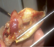
The material was studied in the laboratory of “Antiparasitic Biotechnology” of the department “Biological Safety” of the Kazakh National Agrarian University. The object of research was trout (Fig. 1).
Classical, pathoanatomical methods were used with the preparation of macro and micro preparations from the selected material, histological preparations. Pathohistological examination of organs and tissues was carried out to detect microscopic changes, as well as to study the correlation of internal organs in the application of food.
The material for histological examination was fixed in a 10 % neutral formalin solution. Serial paraffin sections 5 and 6 μm thick were prepared. Ultrathin sections were made on a semi-automated microtome HEOTION ERM 3100 (Fig. 2, a) and on a microtome MC-2.
Celloid, paraffin and frozen sections were prepared. Sections stained with conventional and some special histological methods: hematoxylin-eosin on the apparatus, a processor for coloring the sections of Leica No. S4040 / №000000358 (Fig. 2, b). Histological micro-preparations were studied using a binocular microscope MBI-6 under various magnifications (Fig. 2, c).
Results of macroscopic examination before the experiment
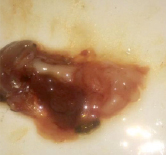
At macroscopic research of a trout: a liver – is increased, yellow color, a consistence flabby. The gallbladder is full of bile orange color. Gastrointestinal tract – pale yellow color. When washed in water, the mucous membrane is swollen, in some places there are spotted hemorrhages. The stomach is empty, shortened, pale gray, spotted hemorrhages were observed in the cardinal part (Fig. 3, a, b).
Swimming bubble. red-yellow color, a testy consistency, a surface on a cut of dark red color, hemorrhages are present under the pleura.
Heart: enlarged in the size of a round-oval shape. Under the epicardium there are many point hemorrhages. Myocardium is red-brown, coronary vessels are slightly filled with blood.
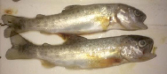
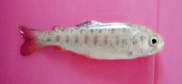
а) b)
Fig. 1. Trout (a) – before the experiment; (b) – after the experiment
|
|
|
|
|
a) Semi-automatic microtome HEOTION ERM 3100 for the preparation of ultra-thin sections |
b) Apparatus-processor for coloring of the Leica incisors No. S4040 / № 000000358 |
c) Equipment for microscopic research and fixation of results |
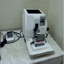
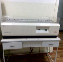

Fig. 2
а) b)
Fig. 3. Gastrointestinal tract and liver of trout
The kidneys are enlarged, swollen, hyperemic, the capsule is easily removed, the cortical layer is dark red, the medulla is red.
Results of macroscopic examination after the experiment
During the experiment using domestic feeds with the inclusion of probiotic preparations, the trout mass increased accordingly, further differentiation occurred in the histological structure.
When examining the material 15 days after the start of the experiment, a noticeable lengthening of the intestine was established (Fig. 4).
Liver – the organ is enlarged, grayish-yellow in color, flabby consistency. The gallbladder is full of bile orange color. Spleen: not enlarged, pulp softened, dark cherry color. Swimming bubble. red color, a testish consistency, a surface on a red cut. Heart – myocardium on a section of uneven color, red-brown areas alternate with light gray dull areas, corresponding to the centers of cardiac muscle dystrophy. The kidneys are slightly enlarged, grayish-yellow in color.
Results of microscopic studies before the experiment
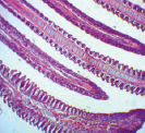
In the histological study of trout gills, the structure of the gill lobes has not been disturbed, the structure of the lamellae is preserved, the epithelial cells are hyperplastic (Fig. 5, a).
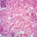
Kidneys – in the cortical layer of the kidney the epithelial cells of the tubules are saturated with eosin, their volume is increased, the walls of the tubules are thickened, the lumens narrowed. The boundaries of nephrocytes are indistinct, not expressed, the nucleus is in a state of pycnosis. Numerous small grains are located in the cytoplasm of cells (Fig. 5, b).
Intestines – the villi are thickened, the apex of many of them is devoid of epithelial lining and merges with the rosy-colored mass of catarrhal exudate that covers the mucous membrane. Crypts (Lieberkunovy glands) are preserved, the cytoplasm of cells is sharply vacuolized in a state of dystrophy mucosa.
At the core of the villi are numerous cell infiltrates: lymphocytes, histiocytes. On the mucosa in the rosy-colored mass of catarrhal exudate, it is possible to see the fallen, decaying cells of the epithelium (Fig. 5, c).
In muscles – the structure of muscle fibers is preserved, the sarcolemma is not broken, in some places the striated striation of the fiber is erased, the number of nuclei is reduced, the intermuscular connective tissue is edematous (Fig. 5, d).
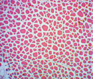
In the liver, the changes were manifested by parenchymal dystrophy of hepatocytes, the vessels in most cases were full-blooded, the endotheliocytes swollen. Hepatocytes are enlarged in volume, the structure of the hepatocyte structure is broken, the number of nuclei is reduced, some nuclei are in the state of pycnosis, rex and lysis. Intermediate and interlobular blood vessels are blood-filled (Fig. 5, e).
Results of microscopic studies after the experiment
Histological examination in gills in trout found that, basically, the structure of lamellae is not broken, the structure of gill lobes is preserved (Fig. 6, a).
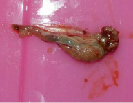
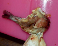
Fig. 4. Gastrointestinal tract of trout at the end of the experiment
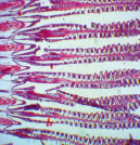
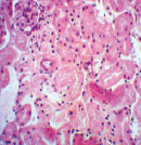
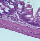
a) Gills b) Kidney c) Intestine
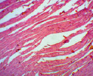
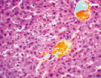
d) Muscle e) The liver
Fig. 5
Kidneys – one canaliculus in the state of granular dystrophy: they are intensively colored with eosin in pink, enlarged in size, their walls thickened, lumens narrowed. The boundaries between the cells are poorly visible. The cytoplasm of cells is fine-grained, the nuclei lie unevenly, some of them in a state of pycnosis and lysis. Other tubules are normal: their lumens are well marked, the nuclei are evenly distributed in each cell (Fig. 6, b).
Histological analysis of the intestine showed that the epithelial cells in the apical part form vacuoles, leading to destruction and destruction of cells. The muscular membrane is thin, the connective tissue tissue is poorly developed, puffiness is observed in it. Outside, all pyloric processes are covered with adipose tissue (Fig. 6, c).
In muscles the structure of muscle fibers is preserved, the sarcolemma is not broken. The nuclei are located on the periphery of the fiber, in places the vacuolization of the intermuscular connective tissue is observed (Fig. 6, d).
In the liver of trout is characterized by an increase in the size of hepatocytes due to the accumulation of lipid inclusions in the cytoplasm. Many hepatocytes were highly vacuolated and had nucleus cells pushed to the periphery. In some hepatocytes, pycnotic nuclei were observed. Histopathological examination of the liver showed that there were changes in many hepatocytes, circular vacuoles corresponding to the locations of the droplets of fat are seen in different sizes. Large vacuoles occupy the entire cytoplasm, so the nucleus is displaced to the periphery of the cell, the cells are filled with one large drop of fat and a nucleus mixed into the periphery. This phenomenon is called cricoid cells (Fig. 6, e).
Conclusion
Thus, on the basis of histological studies, using domestic feeds with the inclusion of probiotic agents, it was established that in the gastrointestinal tract, all experimental trout specimens showed no significant disturbances in the structure of the mucosa and muscle membranes.
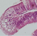
a) Gills b) Kidney c) Intestine
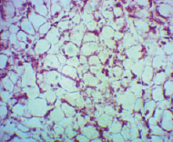
d) Muscle e) The liver
Fig. 6
The revealed changes are compensatory-adaptive.
The histological structure of the liver is characterized by a well-defined tubular structure of the parenchyma with wide sinusoids. The cytoplasm of hepatocytes is homogeneous, but there are individual hepatocytes with small fat vacuoles. Some individuals have vasodilatation and local expansion of sinusoids. In general, the liver of fish has no noticeable pathological disorders.
Pathological changes in muscle tissue and the rest in the organs are considered as moderate.
On the basis of the obtained pathomorphological data, it can be concluded that the positive effects of domestic fodders with the inclusion of probiotic agents, which were characterized by an increase in the mass of individuals, lengthening of the intestine, accumulation of fat and other positive signs. The established minor changes do not tend to disrupt the structure of organs and tissues, do not hinder the growth of fish.
Библиографическая ссылка
Ibazhanova A., Shabdarbaeva G., Kenzhebekova Zh., Khussainov D., Batanova Zh., Tulemisova Zh., Amirgalieva S. HISTOLOGICAL ESTIMATION OF THE STATE OF FISHING MATERIAL AND COMMODITY FOOD PRODUCTS BY APPLICATION OF PROBIOTIC ACTING PREPARATIONS // European Journal of Natural History. 2018. № 2. ;URL: https://world-science.ru/en/article/view?id=33847 (дата обращения: 15.07.2026).