
Scientific journal
European Journal of Natural History
ISSN 2073-4972
ИФ РИНЦ = 0.204
THE INITIAL STAGES OF DEVELOPMENT OF LYMPH NODE CAPSULE IN HUMAN

The structural basis for lymph flow has been a subject of numerous studies, but mostly performed on humans and different mammals lymphatic vessels. Lymph nodes are investigated usually as lymphoid organes [1]. But their capsules contain muscle cells, which provide the energy of phasic contractions to propel lymph downstream towards the central veins as muscle cuff of lymphoid lymphangion [2]. The prenatal development of human lymphatic system did not attract enough attention of the lymphatic researchers due to the understandable difficulties to work with human tissues. E.Klein [4] described their origin from fat tissue. F. Sabin [5] and O. Kampmeier [6] marked that lymphoid tissue appears in walls and partitions of lymphatic sacs from mesenchyme. O. Hertwig [7] described lymph node anlage in human embryos of first months as plexuses of prymordial blood and lymphatic vessels with many mesenchymal cells in their loops. They transform into reticulate and lymphoid cells which isolate as compact mass. On periphery of lymph nodes afferent lymphatic vessels and marginal sinus are formed from lymphatic capillaries. P. Fusardi [8] considered that marginal sinus of the node appears at first after its capsule and parenchyma. According electrone microscope data of R. Markgraf et al. [9], development of human lymph nodes begins from mesenchyme condensation in foetuses of 12-14 weeks. Such anlage consists of mesenchymal cells and capillaries. It invaginates in lymphatic sac lumen. A.Landsberger [10] discerned few fases in development of lymph nodes: anlage - сompact condencations of mesenchymal cells near wall of one or between walls of two lymphatic vessels; primary differentiation - lymph node anlage invaginates in lymphatic vessel lumen, capsule, marginal sinus and parenchyme are formed; secondary differentiation - appearing of lymphoid nodules and medullary sinuses. R. Baily and L. Weiss [11] discovered invaginations of connective tissue with endothelial covering in lymphatic sac of human embryos 30‑37 mm lengths with its dividing into net of spaces. The invaginations contain reticulate cells, macrofages and blood vessels. In human embryos 43-48 mm lengths immature lymph nodes are formed. In their strome lymphocytes are accumulated. In human embryos more 75 mm length mature lymph nodes are appeared - capsule differentiates, ammount of lymphocytes increases. Even existence of muscular structures in capsule of lymph node is discussed in the literature and their development didn t is described.
Material and methods
The work was carried out on 400 both sexes human embryos and foetuses of 4-36 weeks old without pathology. Material was fixed in 10% solution of neutral formalin. Part of material was stained in paraffin with following production of serial longitudinal and transverse sections of 5-10 mkm in thickness. Sections were stained by hematoxylin and eosin, picrofuxine, azane, silver nitrate, orseinum. Lymph node capsules´ total preparations from some foetuses of 19‑36 weeks were stained by gallocyanin.
Results
Anlage of lymph nodes could be determined in human embryos of 28-30 mm length (the ending of week 8): during the dilatation of the paired jugular lymphatic sac the adjoining blood vessels including external coat in their thick walls invaginate into sac s lumen together with its thin endothelial wall and intermediate connective tissue. In result sac´s lumen around the inaginate (stromal anlage of lymph node) narrows and becomes twisted (primary marginal sinus of the node). This process extends on all regions of human foetuses of third month (Fig. 1). Increasing anlage of lymph node is founded on the path of lymph flow and acumulates fragments of degenerating embryonic structures bringing with lymph. Possibly therefore macrofages and lymphocytes migrate from the blood microvessels into stromal anlage of lymph node which becomes lymphoid anlage (Fig. 2). Thus basic lymph collector is divided on the marginal sinuses of lymph nodes and their afferent and efferent lymphatic vessels. Between them could be found border valves. The capsule of lymph node is formed such as walls of its connected lymphatic vessels. Primary capsule represented as very thin adventitial layer covering endothelium of primary marginal sinus in foetuses of 3-4 months contains network of thin reticular fibers. During the month 4-5 the thickness of the capsule increases and its adventitial layer could be divided on two parts: the thin subendothelial layer, which is full of thin reticular fibers, and the thicker outer layer, which is rich by blood capillaries, thick reticular and collagen fibers. The muscle cells are mostly located in between of subendothelial and outer adventitial layer of the capsule (Fig. 3, 4). In this stage of development branches of the capsule and marginal sinus grow into lymphoid parenchyma of lymph node with formation of thin trabeculae and intermediate sinuses. Thickness of the capsule increases especially around blood vessels in the base of the invaginate (hilum of lymph node). In external part of lymph node parenchyma lymphocytes form clots - the primary lymphoid nodules. During month 5-6 the layer of myocytes is still interrupted, and mostly single myocytes or small groups of them could be determined in the capsule. However these cells are getting bigger and denser, particularly in hilum thickerning of the capsule and around mouths of the afferent lymphatic vessels. Together with thin elastic fibers the myocytes form a thin medium layer of the capsule. Thick outer layer of the capsule consists of bundles of thick collagen fibers and elastic fibers. In this stage of human development the lymph node parenchyma divides on the cortex and medulla with cortical and medullary sinuses. Their endothelium extends very much and becomes very thin and perforating. All structures of the capsule including muscle bundles continue in the walls of afferent and efferent lymphatic vessels of lymph node. In foetuses of months 7-9 single myocytes could be determined in external layer of capsule and trabeculae of lymph node. Thus is planned formation of the multi-layered capsule.
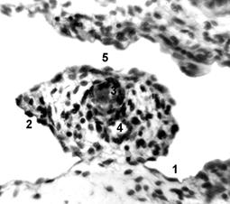
Figure 1. Human foetus week 10.5, cross section: 1 - endothelial wall of posterior pancreaticoduodenal lymphatic vessel; 2 - endothelial cover of stromal anlage of pancreaticoduodenal lymph node; 3,4 - arteriola and venule; 5 - primary marginal sinus of lymph node. Hematoxylin / eosin staining. Light microscopy, magnification - 400X.
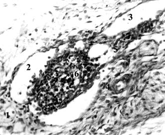
Figure 2. Human foetus week 13, cross section: 1,3 - afferent and efferent lymphatic vessels of pancreaticoduodenal lymph node; 2 - its primary marginal sinus; 4,5 - arteriola and venule; 6 - lymphoid anlage of lymph node. Hematoxylin / eosin staining. Light microscopy, magnification - 240X.
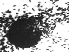
A B
Figure 3. Human foetus week 22, total praparetions of lymph node capsules, superior mesenteric (A) and lumbar (B). Arrows are shown myocytes in the lymph node capsule and in the walls of its lymphatic vessel. Gallocyanin. Light microscopy, magnification - 400X.
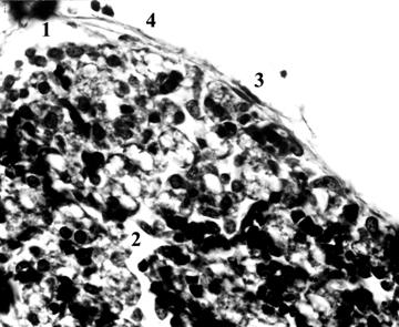
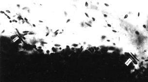
Figure 4. Lumbar lymph node of human foetus week 29, cross section: 1,2 - marginal (subcapsular) and cortical sinuses; 3,4 - bundles of smooth myocytes and elastic fibers in capsule. Orseinum. Light microscopy, magnification - 600X.
Conclusion
The lymph nodes arise from embryonic lymphatic collectors including sacs which form together with blood vessels complexes with intimate connections from ending of human embryogenesis. From anlage lymph node develops in intimate connection with its afferent and efferent lymphatic vessels moreover the inner structure of the node capsule develops such as vessels walls [3]. The capsule diferentiates as thin layer of loose connective tissue with thin reticular fibers covering endothelium of primary marginal sinus in foetuses of 3-4 months. The structural basis of the active lymph transport - muscle cells and valves - could be found in the lymph node capsule already during the first half of the foetal development in humans. At the beginning the valves are appeared, the muscle cells - later. Valves could be found only on entrance in subcapsular sinus of lymph node and on exit of its hilum sinus. Muscular bundles pass from the wall of lymphatic vessels into lymph node capsule and to the contrary. During the second half of the development the maturation of these basic elements of lymphatic pumping is underway - the enlargement of muscle cells together with increases in their quantity leads to formation of the multi-layered capsule of lymph node with more and more thick muscle net in the middle layer.
REFERENCES
- Oord J.J. The immune response in the human lymph node. A morphological, enzyme and immunohistochemical study. Leuven, Kath. Univ. 1985.
- Petrenko V.M. // European Journal of natural history. 2006; 2:134-136.
- Petrenko V.M. // European Journal of natural history. 2009; 6: 25-28.
- Klein E. The anatomy of the lymphatic system. London. 1875.
- Sabin F.R. // Amer.J.Anat. 1909; 9: 43-91.
- Kampmeier O.F. Evolution and comparative morphology of lymphatic system. Springfield, C.Thomas. 1969.
- Hertwig O. Lehrbuch der Entwicklungsgeschichte. 10 Aufl. 1920.
- Fusardi P. // Bull. Ass. Anat. Naney. 1965; 125: 605-618.
- Markgraf R., GaudeckerB., Müller-Hermelink H.K. // Cell Tiss. Ress. 1982; 225 (2): 387-413.
- Landsberger A. // Anat. Anz. 1968; 21: 187-195.
- Baily R.P. a. Weiss L. // Amer. J. Anat. 1975; 142 (1): 15-28.
Библиографическая ссылка
Petrenko V THE INITIAL STAGES OF DEVELOPMENT OF LYMPH NODE CAPSULE IN HUMAN // European Journal of Natural History. 2010. № 3. ;URL: https://world-science.ru/en/article/view?id=21003 (дата обращения: 02.07.2026).