

Genetically determined eye pathologies in the canine population represent a large group of hereditary diseases that significantly reduce vision and quality of life, respectively. In turn, the existing diversity of genetic mutations has a significant impact on the emergence of nosologies specific to individual breeds or breed groups. Collie eye anomaly (CEA) is diagnosed mainly in Collies and related breeds such as Shetland Shepherd, Australian Shepherd, Nova Scotia Duck Tolling Retriever, English Shepherd, etc. [1,2]. Bilateral blindness is quite rare due to the predominantly asymmetrical nature of the lesion and the unequal severity of the process. However, secondary retinal detachment and intraocular hemorrhage significantly worsen the course of the disease [1]. In previously published works, the frequency of occurrence of the pathology varies depending on the country, so among long-haired collies the disease was recorded in the Netherlands in 40.6% of dogs (Stades & Barnett, 1981), in Thailand - 83.3%, and according to data from the University of Veterinary Medicine and Pharmacy in Kosice, Slovakia - 55.5% [1,3]. In the Sheltie, Australian Shepherd, and Border Collie breeds, the incidence is significantly lower—15.1%, 9.71%, and 11.1%, respectively [1,4]. In Australia, in the Australian Shepherd population, collie eye anomaly is the second most common inherited eye disease (Munyard, 2007). Hereditary cataract (РHC) is a progressive clouding of the lens and is the most common cause of vision impairment and loss in dogs. Unlike most susceptible breeds, the Australian Shepherd has an autosomal dominant inheritance pattern with incomplete penetrance [4]. In 12 European countries, as well as in the USA and New Zealand, Australian Shepherds homozygous for hereditary cataract disease were diagnosed in 3.46 to 17.6% of cases, while heterozygous for the mutant allele was diagnosed in 11.2 to 25.5% [4,5]. In general, the nosology is registered in almost 100 dog breeds and can presumably be quite widespread [6,7,8]. Cavalier King Charles Spaniels are particularly susceptible to сongenital keratoconjunctivitis sicca and ichthyosiform dermatosis (CKCSID), a condition characterized by decreased corneal hydration due to lacrimal gland dysfunction. It was first described by Barnett in 2006. According to data provided by the British Trust for Animal Health, in 2012, 0.4% of dogs in the Cavalier King Charles Spaniel breed were homozygous and 10.8% were heterozygous for the disease (Penderis, 2012). Primary lens luxation (PLL), reported in 44 dog breeds, is characterized by hereditary degeneration and destruction of the fibers of the zonular ligament, which holds the lens on the ciliary body within the visual axis, preventing its displacement (Curtis, 1990; Mellersh, 2014). In case of dislocation, the lens is displaced into the anterior chamber of the eye, which most often leads to the development of glaucoma and subsequently to loss of vision (Grahn, 2003; Morris & Dubielzig, 2005; Johnsen, 2006). In some breeds, the pathology has a high degree of heritability, in connection with which DNA testing of animals used in breeding is certainly relevant (Oberbauer, 2008; Betschart, 2014; Gharahkhani, 2015). Coat colour, albinism, oculocutaneous type IV (OCA4 L) is specific to breeds such as the French Bulldog, Pug, Lhasa Apso, Pekingese, and German Toy Spitz. A characteristic feature of the pathogenesis of the disease is a violation of the biosynthesis of melanin and, as a consequence, a decrease or absence of pigment in the skin, hair and eye structures [9]. In addition to the reduced quality of life caused by impaired visual function and increased sensitivity to ultraviolet radiation, the pathology contributes to the development of malignant skin neoplasms [10]. Multifocal retinopathy type 1 (CMR1) is an inherited degenerative disease of the retina and pigment layer caused by a mutation in the BEST1 gene (Guziewicz, 2007; Zangerl, 2010; Hoffmann, 2012). The nosology is registered in at least 12 dog breeds. The disease manifests in puppies at an early age and progresses until 6–12 months of age (Grahn, 2008). In mild cases, the pathology does not cause clinical symptoms and has virtually no impact on the quality of life of the animal and the owner, but there are known cases characterized by severe retinal damage and complete loss of vision [11]. In certain breeds, the incidence and course of the disease may differ [4]. Progressive retinal atrophy (PRA) is the largest group of inherited diseases of the inner lining of the eye, with a wide prevalence among dogs, regardless of country or continent. In modern understanding, more than 100 known dog breeds are susceptible to hereditary progressive retinal atrophy [12,13]. Due to the diversity of mutations in one breed, several types are often diagnosed. The incidence of breed-specific types of progressive retinal atrophy can reach 25% of dogs in the population [14]. The pathology is characterized by vascular degeneration and retinal detachment, loss of pigmentation, increased reflectivity of the tapetum, optic nerve atrophy and, as a result, vision loss [15]. As can be seen from the above, data on the prevalence of most hereditary eye diseases in dogs, taking into account breed specificity, are often absent from published works or have not been updated for a long time. For this reason, an objective assessment of the degree of risk of the emergence and development of individual nosologies in the population is not possible.
The aim of the study is to determine the prevalence of genetically determined eye pathologies in dogs of certain breeds.
Research materials and methods
Retrospective analysis of the results of 18953 molecular genetic examinations in 29 dog breeds for 18 hereditary eye diseases. DNA testing was carried out in the laboratory of the Center for Veterinary Genetics ZOOGEN, St. Petersburg, Russia. The biomaterial was obtained over a period of 15 years (2011–2026) from dogs used in breeding, living in various regions of the Russian Federation. Buccal epithelium or dried blood were used as biomaterial. DNA diagnostics were performed using methods accepted for genetic research: polymerase chain reaction (PCR) followed by electrophoresis, real-time PCR, multiplex PCR, and others.
The results of the study and their discussion
The largest number of studies were performed on the diseases progressive retinal atrophy - 62.54% (n=11854), collie eye anomaly - 15.01% (n=2846) and hereditary cataract - 10.73% (n=2034). Congenital keratoconjunctivitis sicca and ichthyosiform dermatosis, multifocal retinopathy type 1, primary lens luxation and сoat colour, albinism, oculocutaneous type IV were diagnosed in less than 5% of each nosology (Figure).
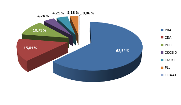
Number of studies performed on nosologies
(PRA-Progressive retinal atrophy, CEA-Collie eye Anomaly, РНS-Hereditary cataract, CKCSID- Congenital keratoconjunctivitis sicca and ichthyosiform dermatosis, CMR1- Multifocal retinopathy type 1, PLL-Primary lens luxation, OCA4 L- Coat colour, albinism, oculocutaneous type IV)
According to the obtained results, breed-specific studies of progressive retinal atrophy are in demand. Thus, among Papillons (pap-PRA) 2662 (22.45%) examinations were carried out, in the population of Golden Retrievers (GR-PRA1/GR-PRA2) - 2414 (20.36%), in five colors of Miniature Schnauzer (PRA-B1) - 2173 (18.33%), Basenji (bas-PRA) - 1367 (11.53%). The types of progressive retinal atrophy characteristic of several dog breeds are somewhat less frequently diagnosed (Table).
Prevalence of hereditary eye pathology depending on the nosology and breed of dogs
|
№ |
Nosology |
Breed |
MM
|
NM
|
NN
|
Number of animals |
|
1 |
Progressive retinal atrophy prcd |
Australian Shepherd |
0 |
0 |
271 |
271 |
|
Chinese Crested Dog |
1 |
20 |
346 |
367 |
||
|
2 |
Progressive retinal atrophy PRA-cord1/crd4-PRA |
American bully |
24 |
88 |
94 |
206 |
|
English Springer Spaniel |
3 |
26 |
48 |
77 |
||
|
Dachshund rabbit smooth-haired |
12 |
47 |
55 |
114 |
||
|
Dachshund rabbit long-haired |
12 |
108 |
71 |
191 |
||
|
Dachshund rabbit wire-haired |
2 |
1 |
18 |
21 |
||
|
Dachshund miniature smooth-haired |
24 |
133 |
106 |
263 |
||
|
Dachshund miniature long-haired |
40 |
338 |
273 |
651 |
||
|
Dachshund miniature wire-haired |
1 |
20 |
28 |
49 |
||
|
Dachshund standard |
0 |
2 |
7 |
9 |
||
|
Dachshund standard long-haired |
0 |
1 |
4 |
5 |
||
|
Dachshund standard wire-haired |
0 |
2 |
7 |
9 |
||
|
French Bulldog |
5 |
20 |
41 |
66 |
||
|
3 |
Progressive retinal atrophy crd1-PRA |
American bully |
4 |
22 |
128 |
154 |
|
American Staffordshire Terrier |
0 |
0 |
235 |
235 |
||
|
4 |
Progressive retinal atrophy crd2-PRA |
American Pit Bull Terrier |
1 |
65 |
301 |
367 |
|
American Staffordshire Terrier |
0 |
1 |
2 |
3 |
||
|
5 |
Progressive retinal atrophy rcd3 |
Chinese Crested Dog |
1 |
0 |
47 |
48 |
|
Tibetan Terrier |
0 |
0 |
4 |
4 |
||
|
6 |
Progressive retinal atrophy rcd4 |
Tibetan Terrier |
0 |
15 |
40 |
55 |
|
7 |
Basenji progressive retinal atrophy |
Basenji |
14 |
350 |
1003 |
1367 |
|
8 |
Progressive retinal atrophy GR-PRA1 |
Golden Retriever |
12 |
190 |
973 |
1175 |
|
9 |
Progressive retinal atrophy GR-PRA2 |
Golden Retriever |
8 |
403 |
828 |
1239 |
|
10 |
Progressive retinal atrophy of papillons and phalens |
Papillon |
15 |
578 |
2069 |
2662 |
|
11 |
Progressive retinal atrophy type B1
|
Miniature Schnauzer black |
1 |
66 |
569 |
636 |
|
Miniature Schnauzer black & silver |
4 |
138 |
665 |
807 |
||
|
Miniature Schnauzer pepper & salt |
1 |
28 |
477 |
506 |
||
|
Miniature Schnauzer white |
0 |
27 |
165 |
192 |
||
|
Miniature Schnauzer chocolate-colored |
0 |
15 |
17 |
32 |
||
|
12 |
Progressive retinal atrophy CNGA1 |
Sheltie |
0 |
0 |
73 |
73 |
|
13 |
Hereditary cataract
|
Australian Shepherd |
8 |
155 |
377 |
540 |
|
Staffordshire Bull Terrier |
0 |
8 |
500 |
508 |
||
|
French Bulldog |
1 |
28 |
957 |
986 |
||
|
14 |
Collie eye Anomaly
|
Australian Shepherd |
8 |
132 |
591 |
731 |
|
Border Collie |
25 |
186 |
444 |
655 |
||
|
Collie long-haired |
213 |
30 |
6 |
249 |
||
|
Sheltie |
401 |
593 |
217 |
1211 |
||
|
15 |
Congenital keratoconjunctivitis sicca and ichthyosiform dermatosis |
Cavalier King Charles Spaniel |
0 |
121 |
684 |
805 |
|
16 |
Primary lens luxation |
Chinese Crested Dog |
19 |
185 |
364 |
568 |
|
Tibetan Terrier |
0 |
3 |
32 |
35 |
||
|
17 |
Coat colour, albinism, oculocutaneous type IV |
French Bulldog |
0 |
5 |
7 |
12 |
|
18 |
Type 1 Multifocal retinopathy |
French Bulldog |
0 |
72 |
727 |
799 |
MM – homozygous for the mutation, NM - heterozygous, NN – homozygous for the reference allele
Dogs homozygous for mutations were identified among the following breeds: American Bully - 11.65% (PRA-cord1) and 2.6% (PRA-crd1), American Pit Bull Terrier - 0.27% (PRA-crd2), English Springer Spaniel - 3.9% (PRA-cord1), Basenji - 1.02% (bas-PRA), Golden Retriever - 1.02% (GR-PRA1) and 0.65% (GR-PRA2), Chinese Crested Dog - 0.27% (PRA-prcd) and 2.08% (PRA-rcd3), Papillon - 0.56% (pap-PRA), French Bulldog - 7.58% (PRA-cord1). Heterozygous for mutations in breeds: American Bully - 42.72% (PRA-cord1) and 14.29% (PRA-crd1), American Pit Bull Terrier - 17.71% (PRA-crd2), American Staffordshire Terrier - 33.33 (PRA-crd2), English Springer Spaniel - 33.77% (PRA-cord1), Basenji - 25.6% (bas-PRA), Golden Retriever - 16.17% (GR-PRA1) and 32.53% (GR-PRA2), Chinese Crested Dog - 5.45% (PRA-prcd), Papillon - 21.71% (pap-PRA), Tibetan Terrier - 27.27% (PRA-rcd4), French Bulldog - 30.3% (PRA-cord1). In Dachshunds, progressive retinal atrophy (PRA-cord1/crd4-PRA) was diagnosed taking into account the breed size of the animals and the morphological features of the coat. Homozygous for the mutation were identified: Dachshund rabbit smooth-haired - 10.52%, Dachshund rabbit long-haired - 6.28%, Dachshund rabbit wire-haired - 9.52%, Dachshund miniature smooth-haired - 9.12%, Dachshund miniature long-haired - 6.14% and Dachshund miniature wire-haired - 2.04%. Heterozygous for the mutation: Dachshund rabbit smooth-haired - 41.22%, Dachshund rabbit long-haired - 56.54%, Dachshund rabbit wire-haired - 4.76%, Dachshund miniature smooth-haired - 50.57%, Dachshund miniature long-haired - 51.92%, Dachshund miniature wire-haired - 40.81%, Dachshund standard - 22.22%, Dachshund standard long-haired - 20% and Dachshund standard wire-haired - 22.22%. Depending on their color, Miniature Schnauzers are divided into five separate breeds. The results of the study can reliably note the heterogeneity of the distribution of рrogressive retinal atrophy type B1 depending on color. In black color, 0.15% of animals were homozygous for the mutation and 10.37% were heterozygous. Among the representatives of the black & silver color, 0.49% were diagnosed as homozygous for the mutation, and 17.10% were heterozygous. In the pepper & salt color, one animal was identified as homozygous for the mutation - 0.19%, while heterozygous - 5.53%. In white and chocolate colors, heterozygous rates were 14.06% and 46.87%, respectively. The largest number of dogs homozygous for the collie eye anomaly mutation were diagnosed in the long-haired Collie breeds – 85.54% and the Sheltie – 33.11%. It should be noted that animals heterozygous for the mutant allele are found in almost half of the cases among Shelties (48.96%). Border Collies and Australian Shepherds had the lowest percentage of homozygotes for the mutation at 3.81% and 1.09%, respectively, while carriers were detected in 28.39% and 18.05% of cases in each breed. According to the results of our study, hereditary cataracts are predominantly distributed in the Australian Shepherd population, with homozygous and heterozygous variants identified in 1.48% and 28.70% of dogs, respectively. Breed-specific сongenital keratoconjunctivitis sicca and ichthyosiform dermatosis in Cavalier King Charles Spaniels was diagnosed in 15.03% of "healthy - carrier" animals. Homozygous for the pathology of primary lens luxation in the Chinese Crested dog breed were identified at 3.34%, heterozygous for the mutation – 32.57%. Among Tibetan terriers, heterozygotes for primary lens luxation were identified at 8.57%. Multifocal retinopathy type 1, which is characteristic of 12 dog breeds, is not widespread among French bulldogs, according to the results obtained, with a heterozygous rate of 9.01%. The smallest number of studies in this breed (n = 9) have been conducted on oculocutaneous albinism type 4, with heterozygotes for the mutation diagnosed in 41.67% of cases. However, the sample size is insufficient for objective conclusions.
Conclusions
The greatest number of studies are performed on diseases such as progressive retinal atrophy, collie eye anomaly, and hereditary cataracts. Breed-specific рrogressive retinal atrophy studies are in high demand, as demonstrated by breeds such as the Papillon, Golden Retriever, Miniature Schnauzer, and Basenji. Less commonly diagnosed types are those specific to several dog breeds. Depending on the color, size of the animals and morphological features of the coat, the incidence of the disease varies within a breed or breed group. Homozygous individuals for the сollie eye аnomaly mutation were found primarily in the long-haired Collie and Sheltie breeds. Heterozygous dogs for the mutation are found in half of the cases among Sheltie representatives. Border Collies and Australian Shepherds showed the lowest percentage of sufferers. Hereditary cataracts, according to our study, are predominantly common in the Australian Shepherd population. Congenital keratoconjunctivitis sicca and ichthyosiform dermatosis, specific to Cavalier King Charles Spaniels, was diagnosed in 15.03% of dogs heterozygous for the mutant allele, which is slightly higher than the results presented by the British Trust for Animal Health. In the Chinese Crested dog breed, every third representative can be “healthy – carrier” of primary lens luxation.
The results of our study confirm a fairly high frequency of occurrence of both homozygous and heterozygous animals for hereditary eye pathologies among the tested dogs. The widespread prevalence of hereditary diseases could be mitigated through screening genetic testing of dogs used for breeding.
Конфликт интересов
Финансирование
Библиографическая ссылка
Горинский В.И. РАСПРОСТРАНЕННОСТЬ НАСЛЕДСТВЕННЫХ ЗАБОЛЕВАНИЙ ГЛАЗ СРЕДИ ОБСЛЕДОВАННЫХ СОБАК // European Journal of Natural History. 2026. № 1. ;URL: https://world-science.ru/ru/article/view?id=34427 (дата обращения: 05.09.2026).
DOI: https://doi.org/10.17513/ejnh.34427