

In recent years World healthcare organization sees widespread of obesity as an epidemy that covers all countries and continents of our planet. Obesity causes many negative consequences in our organism, apart from damage of cardiovascular system and motor apparatus, development of sugar diabetes, specialists name complications of pregnancy and delivery, and also high frequency of antenatal damage of embryo. All these factors considered, obesity among women of fertile age group became a serious problem of modern obstetrics [1, 2, 3]. Regardless of many researches, devoted to this problem, mechanisms of placental insufficiency formation, as well as aspects of placental structure among pregnant women who suffer from obesity, and dependence of structural – functional disturbance in placenta upon obesity degree are not studied sufficiently.
Modern concepts on mechanisms of development of placental insufficiency is based upon evaluation of compensatory-adaptive reactions of placental complex [4]. The main pathogenetic mechanism in development of placental insufficiency in case of obesity of mother is considered disfunction of endothelium that causes circulatory disturbance and structural changes in vessel wall which results in decrease in blood flow speed and tissue edema and also sclerotic processes [5]. As a result, both mass and volume of placenta decreases, functional insufficiency of organ decreases [6]. Lack of a single opinion on understanding pathogenetic mechanisms of placental insufficiency among women with obesity defines the urgency of studying structural-functional foundation of placental insufficiency in case of presence of this pathology in comparison to placenta of women with normal body mass.
Objective: study morphological structure of placenta among women who suffer from obesity of different degree.
Materials and research methods
The research included 224 placentas of women who suffer from obesity and delivered alive infants in a full-term period. Diagnosis of obesity was established according to definition of index of body mass (IBM). Placentas were divided into 4 groups. Three experimental groups: group I – placentas of women with obesity of the 1st degree (IBM 31,88 ± 1,4kg/m2), group II – placentas of women with obesity of the 2nd degree (IBM 36,6 ± 1,07kg/m2), III – placentas of women with obesity of the 3rd degree (IBM 42,2 ± 1,9kg/m2) (р ≤ 0,05). Control group was formed of placentas of women with normal IBM (21 ± 1,88 kg/m2). Average age of women in group 1 – 30,3 ± 0,5, in group 2 – 30,0 ± 0,9, in group 3 – 33,5 ± 0,8. Average age of women of the control group equaled 28,5 ± 0,7. In all four groups biparas prevailed.
The research excluded women with chronic arterial hypertension, sugar diabetes, preeclampsia, and other vascular extragenital pathology.
Samples of placenta were collected through the whole layer of organ: in central, paracentral, and edge areas of placenta. Fragments were fixed in 10 % solution of neutral buffered formalin, processed according to the standard in series of spirits of an increasing concentration, and sealed in paraffin. Histologic cuts were colored with hematoxilin and eosin. For visualization microscope Leica-1000 with digital photocamera was used.
Observative light-optical microscopia of placentas was carried out as well as morphometry of structural components according to the method by G.G. Avtandilov [7]. It allowed us to evaluate condition of vessel bed of terminal hairs, density of symplastic knots, formation of syncitial-capillary membranes. For this purpose structure of hair tree was determined with micro-preparations: average number and quantitative density of hairs (Nai), volumetric density of terminal hairs (Vv), volumetric density of symplastic sprouts (Vv), volumetric density of free cytotrophoblast (Vv), volumetric density of vesselless hairs (Vv), average number and quantitative density of hair capillars (Nai), volumetric density of syncitial-capillary membranes (Vv).
Statistical procession of data was carried out with programme Statistica7.0 for Windows. To evaluate differences between data T-criterion of Student was used. Difference between indexes of the corresponding groups was considered significant for р ≤ 0,05.
Research results and discussion
According to majority of authors, women with obesity often give birth to children with macrosomia [8]. However, statistically-reliable difference in values of average mass of embryo in experimental groups, compared to the control group, was not registered within our research (table 1).
According to bibliographic data, display of placental insufficiency are mainly related to deficit of endothelial factors (prostacyclin, nitrogen oxide, etc.) that enable relaxation of placenta vessels in normal conditions. Decrease in influence of factors of endothelial relaxation can cause spasm in vessels of feto-placental complex, as a result, circulation disturbance and hypoxia, related to decrease in blood flow speed, develops in placenta, edema and sclerotic processes develop [9]. According to O.B. Karelina [6], both mass and volume of placenta decreases among women with high IBM. Results of our research confirm this statement to a certain degree: difference in mass of placenta between groups of patients with obesity of I-III degree is not as significant as between placenta mass of patients with increased IBM, compared to the control group: thus, average mass of placenta for obesity of the 1st degree was on average 20 % bigger than for obesity of the 3rd degree, while difference between placentas of patients with obesity of the 3rd degree and the control group was about 34 % (table 1).
While studying structural organization of placenta among pregnant women with obesity, we revealed a statistically-significant decrease in volumetric density of terminal hairs depending on increase in degree of obesity (for group I – multiplied by 3,7, for group II – by 4,8, for group III – by 5 in comparison to value of the control group). A progressing decrease in number and quantitative density of capillars in terminal hairs of placenta among women with obesity was established: in group I – by 2, in group II – by 2,2, in group III – by 2,5, compared to value in the control group. The received result allow us to suggest existence of a different, not described before, and, possibly, the leading morphological substrate in development of placental insufficiency in terms of obesity – disturbance of vasculogenesis.
It has been established that along with increase in obesity degree suppression of adaptive processes in placenta becomes more expressed: in comparison to the control group a statistically-reliable decrease in volumetric density of chorial symplasts took please depending on degree of obesity: for group I – by 1,5, for group II – by 2,2, for group III – by 2,4. In case of obesity among the pregnant volumetric density of syncitial-capillary membranes also decreased: considerably in comparison to the control group (for group I – by 4, for group II – by 4,7, for group III – by 5,4), and less significantly – from the 1st group to the 3rd group of obesity (table 2). On the opposite, along with increase in IBM, volumetric density of sclerotic vesselless hairs increases in comparison to the control group: in group I – by 2,5, in group II – by 3,8, in group III – by 4,7 (table 2).
Table 1
Clinical-morphological parameters of feto-placental complex of women with obesity and normal IBM
|
Parameters |
Group I |
Group II |
Group III |
Control group |
|
Mass of placenta (g) |
495,1 ± 10,50* |
457 ± 12,6* |
410 ± 10,8* |
620 ± 12,3 |
|
Placental-embryo coefficient |
0,17 ± 0,025 |
0,16 ± 0,030 |
0,16 ± 0,030 |
0,19 ± 0,030 |
|
Mass of newborn (g) |
3369,3 ± 88,92 |
3245,7 ± 123,91 |
3267,8 ± 98,33 |
3365,5 ± 65,94 |
|
Thickness of placenta (cm) |
2,5 ± 0,17 |
2,5 ± 0,15 |
2,3 ± 0,15 |
2,8 ± 0,17 |
Note: (*) – statistically-reliable difference of the respective indicator in comparison to the control group, р ≤ 0,01.
As we conclude from the received data, in placenta of women with obesity involutive-distrophic changes in different combinations with compensatory-adaptive reactions were registered (plethora of hairs, big number of chorial symplasts, developed syncitial-capillary membranes). We should underline that morphological display of compensatory-adaptive reactions were more expressed in case of obesity of the 1st degree (fig. 1) and decreased along with growth in degree of obesity.
As degree of obesity increases, in placenta, along with compensatory-adaptive processes we registered progressing pathological changes such as fibrinoid necrosis of hairs, fibrinoid mass deposits with immurement of hairs, sclerosis of hairs, decrease in number of syncitial sprouts. These factors are a result of tissue ischemia, and cause decompensated placental insufficiency, formation of fibrinoid clots in inter-hair space, decrease in mass of placenta, and decrease in placental-embryo coefficient. These structural changes in placenta were most typical for group III of placentas (fig. 2).
The progressing suppression of compensatory-adaptive reactions in placentas of women along with increase in degree of obesity is confirmed by the received decrease in area of placental membrane (table 2) that leads to decrease in diffusion of gas and nutrients through placental barier, providing for development of placental insufficiency; interestingly, similar changes in morphological structure of placenta are described in case of moderate and heavy preeclampsia [9].
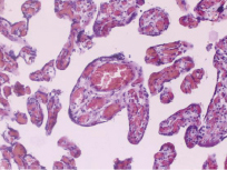
Fig. 1. Plethora of capillars of placenta hairs of a puerpera with obesity of the I degree. Coloring by hematoxilin and eosin; optical multiplication x 200
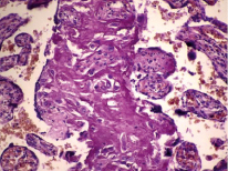
Fig. 2. Deposits of fibrinoid mass with immurement and degeneration of hairs, singly sclerotic hairs in placenta of a puerpera with obesity of the III degree. Coloring by hematoxilin and eosin; optical multiplication x 200
Table 2
Results of morphological research of structural organization of placenta among pregnant women with different degrees of obesity
|
Vv of terminal hairs |
Vv of symplastic knots |
Vv of vesselless hairs |
Nai of capillars |
Vv syncitial-capillary membranes |
|
|
Control group |
68,3 ± 1,07 |
5,4 ± 0,16 |
0,7 ± 0,27 |
14,0 ± 0,42 |
12,1 ± 0,40 |
|
Group I |
18,2 ± 0,72* |
3,72 ± 0,3 |
1,7 ± 0,30* |
7,2 ± 0,40* |
3,0 ± 0,30* |
|
Group II |
14,2 ± 0,71* |
2,4 ± 0,13 |
2,7 ± 0,24* |
6,50 ± 0.30* |
2,55 ± 0,16* |
|
Group III |
13,6 ± 0,6* |
2,2 ± 0,21 |
3,3 ± 0,41* |
5,54 ± 0,33* |
2,2 ± 0,16* |
Note: (*) – statistically-reliable differences of signs in comparison to the control group, р ≤ 0,01.
Disturbances of vessel bed and hair tree in placentas of women who has excessive body mass, described in this research, were expressed the most in case of the III degree of obesity and, of course – in comparison to placentas of women with normal IBM.
Conclusion
Pathomorphological display of placental insufficiency in case of obesity are defined by disturbance in formation of vessel bed of hairs and is defined by degree of obesity. Formation of placental insufficiency along with increase of obesity degree is attended by decrease in expression of compensatory-adaptive reactions and prevalence of pathological changes of placenta structure.
Библиографическая ссылка
Datsenko N.S., Nikitenko E.V., Ageyeva T.A., Yakimova A.V. MORPHOLOGICAL ASPECTS OF PLACENTA AMONG WOMEN WHO SUFFER FROM OBESITY // European Journal of Natural History. 2020. № 3. ;URL: https://world-science.ru/ru/article/view?id=34076 (дата обращения: 25.06.2026).