

Current increased abrasion classifications fail to meet the systematization of clinical aspects of this pathology [3, 5, 10, 11]. The horizontal form of abrasion is diagnosed accurately, but vertical, mixed and other forms are hardly differentiated. That’s why besides horizontal form we distinguish vertical-distal and vertical-mesial, depending on occlusion and position of temporomandibular joints elements. Gnathic part of the face is the variable structure of the craniofacial complex. Vertical parameters are most prone to changes, due to anatomical and physiological features of head growth and development (dental changes, occlusion abnormalities, loss of teeth, increased tooth abrasion, etc.). The interalveolar height increase can lead to a bigger muscles of mastication tonus, and cause dysfunction of the temporomandibular joints and structural changes in the jaw bone [4, 5, 8, 10, 11].
There are decompensated and compensated IAT. Decompensated is accompanied by a decrease in the gnathic part of the face height, and in case of compensated IAT the gnathic part of the face height does not decrease or its height change is insignificant. This is due to vacant (false, compensative) hypertrophy of the alveolar crests bony structures [3, 4, 8, 9, 10, 11].
The decrease in the gnathic part of the face height is affected not only by the abrasion of the teeth degree, the anomaly of occlusion in various directions, but also by changes in the maxillofacial region that occur with increased abrasion of hard tooth tissues, loss of antagonists and other associated pathological conditions [6, 8, 9]. At the same time, there is no clear distinction between the forms of lowering the gnathic part of the face height within patients with increased teeth abrasion. The basic morphometric parameters of patients with a reduced gnathic part of the face are not shown.
Goal of research
Determination of the basic face morphometric parameters within patients with decompensated vertical-mesial form of increased teeth abrasion.
Matherial and methods of research
Morphometric examination of parameters was carried out within 30 patients (12 men and 18 women) with decompensated vertical-mesial form of increased abrasion of teeth.
The comparison group consisted of 64 people (27 men and 37 women) with physiological occlusion and intact dentition.
Kefalometric measurements were carried out with reference to the instructions of J.J. Roginsky (1968), F.J. Horoshilkina (1991), and in accordance with the requirements of anthropometry, which provide the definition of the distance between conventional points. A standard caliper with a fission rate of 0.01 mm was used as a tool.
10 parameters of the brain and facial region of the head were included to the statistical processing. Morphometric parameters were measured when the head was placed in the oculoauricular (Frankfurt) horizontal.
Following measuring points were used during cephalometry to determine the main parameters of the head and face in different directions (Fig. 1).
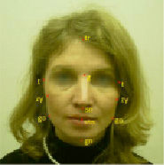
Fig. 1. A photograph of a person with dotted cephalometric points (frontal plane)
Glabella – glabella (g) – the most protruding forward point on the midline between the eyebrows;
Trichion – trichion (tr) – the upper point of the median line of the forehead on the border of the scalp;
Chin – gnathion (gn) – a point located on the middle line on the lower edge of the chin;
Upper nasal – nasal (n) – a point located above the root of the nose at the level of the nasophilic suture;
Subnasal – subnasale (sn) – a point located at the intersection of the nasal septum with the upper lip;
Oral – stomion (sto) – the point of intersection of the line of closure of the lips with the mid sagittal plane;
Zygomatic – zygion (zy) – the most prominent point outside the zygomatic arch;
Goat – tragion (t) – the point located on the upper edge of the tragus of the ear;
Mandibular – gonion (go) is the most outward point on the corner of the lower jaw;
Pogonion – pogonion (pg) – the most protruding point on the chin.
Distances between individual cephalometric points made it possible to determine the size of the head and face in different directions and to evaluate the parameters of the gnathic part of the face with physiological occlusion of permanent teeth. Measurements on the head and face were carried out in three mutually perpendicular directions.
In the sagittal direction, the length of the head (g-ops), the distance from the tragus of the ear to the nasal points and points on the lower jaw (t-n; t-sn; t-pog; t-gn). The depth of the face was calculated with the formula using distances between points (t – t) and (t – n).
Results of the research and their discussion
Law of the relation between the shape of the head and various morphometric parameters of the face is established within the examined control group.
We have determined the morphological features of the head structure and its separate parts (face, gnatical part of the face, intergnatic distance, lower jaw). Morphometric parameters are considered in accordance with sexual dimorphism. The sizes of the head and face were studied. The relation between individual morphometric parameters of the head and face in men and women were determined.
The study results of the morphometric parameters of the head with comparison group are presented in the table 1.
Table 1
Morphometric parameters of the head and face
|
Morphometric parameters |
Face size (mm) |
|
|
males |
females |
|
|
n-mе (face height) |
125,64 ± 6,3 |
111,87 ± 2,26 |
|
gl – me |
136,75 ± 3,29 |
122,34 ± 2,34 |
|
n-inc (height of nasomaxillar complex) |
81,47 ± 3,52 |
73,45 ± 2,24 |
|
sn-inc (height of dental alveolar part of upper jaw) |
21,82 ± 1,17 |
20,07 ± 1,24 |
|
n-sn |
61,19 ± 2,7 |
58,57 ± 2,29 |
|
sn-spm (intergnathic height) |
55,84 ± 4,5 |
53,16 ± 1,54 |
|
gn – me |
6,52 ± 1,29 |
6,02 ± 1,19 |
|
inc – spm (height of dental alveolar part of lower jaw) |
21,96 ± 1,89 |
21,14 ± 1,27 |
|
zy – zy |
143,57 ± 5,1 |
138,41 ± 3,72 |
|
sn – gn |
65,46 ± 1,43 |
63,28 ± 2,16 |
Table 2
Morphometric parameters of the facial skeleton measurement within patients with decompensated vertical-mesial form with increased abrasion of teeth
|
Morphometric parameters |
Face size (mm) |
|
|
males |
males |
|
|
n-mе (face height) |
108,6 ± 3,11 |
105,26 ± 2,53 |
|
gl – me |
118,3 ± 3,29 |
115,24 ± 2,33 |
|
n-inc (height of nasomaxillar complex) |
71,53 ± 2,56 |
69,45 ± 2,34 |
|
sn-inc (height of dental alveolar part of upper jaw) |
12,36 ± 2,37 |
11,6 ± 1,62 |
|
n-sn |
52,18 ± 3,26 |
52,44 ± 3,54 |
|
sn-spm (intergnathic height) |
36,68 ± 2,32 |
34,71 ± 1,47 |
|
gn – me |
43,31 ± 3,65 |
42,51 ± 2,84 |
|
inc – spm (height of dental alveolar part of lower jaw) |
14,96 ± 1,79 |
13,54 ± 1,37 |
|
zy – zy |
133,27 ± 6,35 |
131,471± 6,73 |
|
sn – gn |
54,37 ± 2,16 |
52,56 ± 1,56 |
Thus most of the morphometric parameters of the head and face within male patients were larger than within females and were significantly different.
In general, the morphological height of the face and the height of the nasal part within males were larger than within females. At the same time, the height of the gnathic part was not significantly different in men and women.
The height of the lower jaw (inc- me) was mostly twice the size of the dental alveolar part of both the upper and lower jaws. The joint height of the dental alveolar parts of the upper and lower jaws corresponded to the dimensions of the lower jaw height, and according to these signs of sexual dimorphism was not identified. The height of dental alveolar part of the upper jaw (sn – inc) corresponded to the dental alveolar part of the lower jaw (inc – spm) both in males (21,82 + 1,17 and 21,96 + 1,89 respectively) and females (20,07 + 1,24 and 21,14 + 1,27 respectively).
The results of the facial skeleton measurement within patients with decompensated vertical-mesial form with increased abrasion of teeth are shown in the table 2.
The results of the study showed that the height of the face nasal part (n-sn) corresponded to the lower part of the face (sn-gn) and the difference in these parameters was about 1-2 mm. It should be noted that the height of the dental alveolar part of the upper jaw (sn – inc) was about three times smaller than the height of the nasal part of the face and did not correspond to the dental alveolar part of the lower jaw (inc – spm). The height of the intergnathic part (sn-spm) was reduced by 9-13 mm. Thus, the height decrease of the gnathic part of the face, especially the height of the lower jaw and intergnathic distance, was evident within patients. The interocclusion gap within the patients of this group was between 4-10 mm.
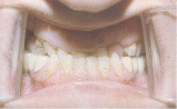
Fig. 2. Oral cavity of the patient Sh. 54 years. The presence of vertical-mesial facets of abrasion of teeth
During the oral cavity examination within patients of this group, there were objected a decrease in the interalveolar height, the presence of defects in the dentition, dental alveolar deformation of the dentition, the facet of abrasion in the vertical-mesial direction (Fig. 2).