

The structural basis for lymph flow has been a subject of numerous studies. Valves divide lymphatic vessels on lymphangions and are considered as passive restrictions of reserve lymph flow, muscular cuffs of lymphangions - as lymphatic pumps [1, 2]. H. Mislin [2], rounding off totals of own and others investigations of lymphangion′s problem during 30 years, whrote about interrupted muscular coat in lymphatic vessel in connection with absent or little few of myocytes about valves. Therefore it is significant that lymphangions make separative, solitary contractions although in the large lymphatic vessels of limbs and in thoracic duct (TD) are discovered cases of combined contractions of 2-4 neighbouring lymphangions. But their morphological bases are not described.
Materials and methods of research
The work was carried out on 12 both sexes dogs of 3-5 years old. TD pick out whithout preliminary injection. Material was fixed in 10 % solution of neutral formalin. Part of material was stained in paraffin with following production of serial longitudinal and transverse sections of 5-7 mkm in thickness. Sections were stained by picrofuxine, azane, total preparations - by gallocyanin.
Results of research and their discussion
TD of dog contains 10-12 valves, constantly - in the ending and supraaortic segments, behind oesophagus (3, more often - 1-2), near aortic arch (2) and diaphragm, above cisterna chyli (3-4), least of all (0-1) - in middle thoracic part (interazygoaortic segment). Valves divide TD on intervalvar segments with different length or lymphangions. TD muscular coat consists of deep (circular) and superficial, subadventitial (longitudinal) layers. Circular muscular bundles have lesser thickening (more often - 1-2 rows of the cells), than longitudinal (to 3-4 rows of the cells), but settle down more tightly on the perimeter of TD. In some places muscular layers become one, particularly in muscular cuff of lymphangion and valvar shaft. In such parts are prevailed oblique bundles with oblique transverse or intermediate (40-50 °) orientation. Intima and adventitia contain dispersed muscular bundles with mainly oblique longitudinal orientation, they not form continuous layers on extent and perimeter of TD or often in one of TD lymphangion. Quite often intimal and adventitial bundles of myocyties anastomose with muscular layers of media or even become in them. In basis of valve circular layer of media thickens and forms projection into valvar shaft, where its oblique transverse bundles interplace with longitudinal muscular bundles of intima, which can change their direction on radial. Subadventitial muscular bundles pass over basis of valve without interruption, but can stretch and thinen. And muscular bundles of intima in basis of valve in some places continue without interruption in proximal direction. Then on section of TD valvar part are discovered two longitudinal muscular layers, separating layer of connective tissue with possible participation of transverse myocyties - marked muscular connections between muscular cuffs more often of first TD lymphangions (fig. 1-3). These longitudinal muscular layers can draw closer to one another in muscular cuff of proximal lymphangion and joint in one layer of media.
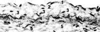
Fig. 1. Thoracic duct of dog, middle part, longitudinal section through muscular cuff: 1 - endothelium (pallid-grey nuclei);
2 - longitudinal myocyties (black nuclei) in intima and internal elastic membrane;
3 - circular myocyties of media;
4 - subadventitial longitudinal myocyties and external elastic membrane;
5 - adventitia and its myocyties. Picrofuxine. Light microscopy, magnification - 300X
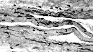
Fig. 2. Thoracic duct of dog, middle part, longitudinal section: 1 - circular myocyties in valvar shaft;
2 - internal elastic membrane and longitudinal muscular bundles of intima, penetrating into valvar cusps and lateral wall of valvar sinus; 3 - subadventitial overvalvar bundle of myocyties and external elastic membrane; 4 - circular muscular layer in lateral wall of valvar sinus. Picrofuxine. Light microscopy, magnification - 300X
а б
в
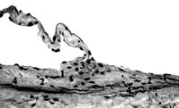
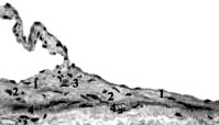
Fig. 3. Thoracic duct of dog, initial part, longitudinal sections: 1 - longitudinal myocyties in intima and internal elastic membrane; 2 - circular myocyties of media; 3 - conjestion of myocyties with different orientations in valvar shaft; 4 - overvalvar (subadventitial) longitudinal muscular bundle and external elastic membrane. Picrofuxine. Light microscopy, magnification - 300X
TD valves are circular folds of its wall, mainly - intima and circular muscular layer. Internal elastic membrane noticeable loses shape in basis of valve: it turns out wall of distal lymphangion in axial sector of valve, become interrupt in parietal sector of valvar shaft (stretching of network of elastic fibres). Between its two layers of intima there is conjestion of myocyties and collagen fibres, which draw closer with parietal intimal layer and thinen in valvar cusp. The most large muscular bundles of intima connect muscular cuff of lymphangion with its entrance valve and lateral walls of valvar sinuses (1) and with its exhaust-valve and lateral walls of axial sinus (2). Longitudinal valvar bundles of myocyties from distal lymphangion continue through shafts and comissurae of border valve into longitudinal commissural bundles of myocyties of proximal lymphangion, or pass by cusps and through lateral (intershaft) walls of axial sinus and valvar comissurae - transvalvar muscular system of neighbouring lymphangions. It can trace the best on the total preparations of TD.
In initial part of TD circular bundles of myocyties settle down into 2-3 layers, limiting TD dilatation and stretching its walls during large throw out of lymph from cisterna chyli. Muscular bundles branch out, intersect and connect in different degree in basic, circular muscular layer of TD, where myocyties settle down tightly, often as continuous muscular stratum. Intima in initial part of TD contains good-marked longitudinal muscular bundles: intensifying transvalvar muscular system, probably, increases resistence of valves in the part to shock pressure of lymph flow from cisterna chyli. Intima in middle thoracic part of TD contains myocyties smaller, circular bundles of its media settle down more crumbly, but subadventitial longitudinal bundles of myocyties are marked powerfully: it can propose more external pressure on TD wall - influence of respiratory excursions of thorax.
Conclusion
Structure of dog TD similar to structure of human TD with cisterna chyli [3]. Valves divide TD on row of intervalvar segments with different length and myocyties in the walls or lymphangions. Muscular coat of TD is the hole, although uneven formation on its extent. Provisionally it can select two muscles - muscular cuff and valvar muscle, but don′t as separate, independent formations, and as local thickening of the muscular coat. These muscles are connected between them as within the lymphangion limits (its muscular cuff and valves), so on the extent of TD - muscular connections of neighbouring lymphangions, transvalvar and overvalvar. Circular bundles of media and longitudinal bundles of intima from muscular cuff of the lymphangion pass through valvar shafts and cusps, uniting the main components of the lymphangion, and form transvalvar muscular system of neighbouring lymphangions. Longitudinal muscular bundles of adventitia (deep layer) and media (superficial, subadventitial layer) pass without interruption over border valves of neighbouring lymphangions and unite their muscular cuffs into one muscular stripe: overvalvar muscular bundles are structural basis for direct spreading (short way) of wave of muscular excitement and combined contractions of neighbouring lymphangions. Since valve is situated under angle to TD wall, wave of muscular excitement passes in it more long way, then by overvalvar muscular bundles. Therefore the work (А) of transvalvar muscular bundles, making (F) during their contractions and directing on axial promotion (s) of lymph, is smaller effective, according to known formula (A = F∙s∙cos α). When valve is turned off (α → 90°), the work become zero, in difference from overvalvar bundles (α → 0°, cos α → 1).
References
- Horstmann E. Uber die funktionelle Struktur der mesenterialen Lymphgefasse // Morphol. Jarb. - 1951. - Bd. 91, № 4. - S. 483-510.
- Mislin H. The Lymphangion // Lymphangiology. - Stuttgart. - New York: Schaffauerverlag, 1983. - P. 165-175.
- Petrenko V.M. Functional morphology of lymphatic vessels (In Russian). Second edition. - SPb, DEAN, 2008. - 400 p.